
In contemporary orthopedic practice, especially within the realm of minimally invasive spine surgery (MISS), the precision and reliability of intraoperative imaging are paramount to achieving optimal surgical outcomes. The advent of the advanced 3D C-arm for minimally invasive spine surgery has revolutionized intraoperative visualization, facilitating unprecedented accuracy in complex spinal interventions.
1. Limitations of Conventional 2D Fluoroscopy in Minimally Invasive Spine Surgery
Traditional two-dimensional (2D) fluoroscopy remains widely utilized; however, it is constrained by inherent limitations, including:
- Superimposition of anatomical structures, resulting in compromised visualization of critical landmarks
- Dependence on subjective interpretation, particularly among orthopedic surgeons with limited radiological expertise
- Inability to obtain accurate linear measurements, due to magnification and distortion, which impedes precise preoperative planning for implant placement
- Challenges in cases of complex anatomy or congenital deformities, such as severe spondylolisthesis or congenital kyphosis, where 2D imaging fails to fully capture spatial relationships
These factors collectively reduce surgical precision and increase the risk of hardware misplacement..
2.Intraoperative Benefits of Advanced 3D C-arm Imaging for Spine Surgery
The advanced 3D C-arm for minimally invasive spine surgery overcomes these limitations by providing:
- Multiplanar reconstruction encompassing axial, sagittal, and coronal views, enabling comprehensive real-time assessment
- Elimination of overlapping structures inherent to 2D imaging, thereby improving anatomical clarity
- High-resolution, high-contrast imaging capable of delineating both osseous and soft tissue structures with remarkable fidelity
- Comprehensive visualization of the entire surgical field throughout the procedure, minimizing intraoperative uncertainty and blind spots
- Immediate intraoperative verification of implant positioning, which is critical for reducing revision rates and enhancing patient safety

3. Key Clinical Applications of Advanced 3D C-arm in Minimally Invasive Spine Surgery Procedures
The clinical integration of advanced 3D C-arm for minimally invasive spine surgery has significantly enhanced surgical accuracy and patient outcomes across a wide range of spinal procedures. One of the most common applications is during thoracic and lumbar pedicle screw fixation, particularly in cases involving unstable fractures or mild spondylolisthesis. Traditional 2D fluoroscopy often struggles with overlapping structures and limited perspectives, whereas 3D imaging offers surgeons a complete spatial view, minimizing errors and improving screw trajectory planning.
In anatomically challenging cases—such as patients with severe spinal degeneration, high-grade spondylolisthesis, or congenital deformities—the benefits of intraoperative 3D visualization become even more critical. The system allows for accurate assessment of pedicle morphology in the cervical and thoracic spine, where standard 2D views may be insufficient or misleading.
Moreover, advanced 3D C-arm systems like the Perlove PLX C7500A and PLX C7600 series are designed to support high-precision imaging during minimally invasive spine surgeries (MISS), providing real-time multi-planar reconstruction that helps reduce surgical invasiveness and improve overall efficiency.
Common MISS applications include:
- Thoracic/lumbar pedicle screw placement
- Cervicothoracic junction surgeries with high anatomical variation
- Complex deformity corrections (e.g., congenital scoliosis, kyphosis)
- Upper cervical instrumentation
- Minimally invasive spinal fusions
Numerous clinical studies support the effectiveness of intraoperative 3D imaging in spinal surgeries. One such study confirms that 3D C-arm–based navigation significantly improves pedicle screw placement accuracy, particularly in complex cases or minimally invasive approaches
👉 Three-Dimensional C-Arm-Based Imaging and Navigation in Minimally Invasive Spine Surgery (NCBI)
4. Technological Trends and Future Directions
The evolution of the advanced 3D C-arm for minimally invasive spine surgery is characterized by:
- Integration with artificial intelligence (AI) for automated anatomical segmentation and surgical planning
- Accelerated 3D image reconstruction algorithms reducing intraoperative downtime
- Development of low-dose imaging protocols to minimize radiation exposure without compromising image quality
- Enhanced interoperability with robot-assisted surgical systems and real-time navigation platforms, fostering the emergence of fully digitalized operating suites
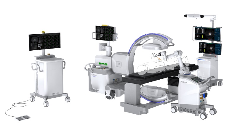
Perlove Medical’s PLX C7500A and PLX C7600 series exemplify these advancements, delivering unparalleled imaging performance and operational flexibility tailored for modern spinal surgery demands.


